Buccal Epithelial Cells with Adherent Bacterial Cells Revealed by Negative Staining
Instructor Version (go to Student Version)
| Subject Area(s) | microbiology |
| Intended Audience |
Middle school science, high school biology, introductory undergraduate microbiology |
| Type | laboratory exercise |
| Revision Date | May 7, 2008 |
INTRODUCTION
The human body represents an enormous and extraordinarily diverse collection of surface habitats and micro-niches potentially available for colonization by biofilm associated micro flora (Denyer et al. 1993). At birth the body is essentially sterile but soon becomes colonized by a variety of microorganisms. With continuous exposure to environmental surfaces and to human contact the biofilms become increasingly complex and gradually approach those typical of adults for a given geographical and perhaps even ethnic population. This accumulation of microbial cells eventually out numbers the human cells in the body by as much as tenfold. Despite this huge population of microbes, much of the body remains sterile in healthy individuals. The blood, and most body fluids, the heart, skeleton and skeletal muscles and the tissues of most organs, brain, liver, kidneys, the lymphatic system and the body cavities remain sterile until exposed to serious illness or injury. Other body regions including the skin, the gastrointestinal tract, the urogenital tract and the oropharynx develop a rich and diverse array of biofilms.
The flora of the mouth is one of the most intensively studied human microbial communities. Over 500 different species of bacteria have been identified in the oral cavity. Microbiologists have concentrated on the microbes that form plaque on the teeth because this flora is responsible for dental caries and periodontal disease (Kolenbrander and Palmer, 2004). Relatively few investigations of the buccal mucosa have been done and those pertain mostly to children (Wilson 2005).
The major colonizers of oral squamous epithelial cells are facultatively anaerobic Streptococci of the viridans group, including S. oralis, S. mitis, S. salivarius and S. sanguis. Other organisms present in abundance are Neisseria spp., Haemophilus spp., Veillonella spp., Lactobacilli, Actinomyces spp. and Propionibacterium spp. (Wilson 2005). In healthy individuals, these are all considered normal flora and not associated with any disease process.
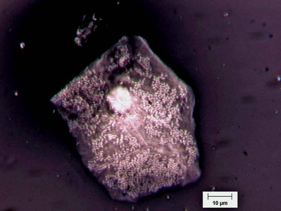
Figure 1 shows a squamous epithelial cell covered with a large number of bacterial cells, most which appear to be diplococci. Wilson (2005) reports a typical number of bacteria per cell between 5 and 25 with this low number being due to the high rate of shedding (desquamation) of epithelial cells. The cell pictured, however, is estimated to have more than 1300 adherent bacterial cells.
PREREQUISITES
Students should be able to define the term biofilm, describe the differences between biofilm (surface-attached) and planktonic (suspended bacterial cells) bacteria, and be able to describe why bacteria usually grow on surfaces. They should understand the distinction between prokaryotic and eukaryotic cells. It is also important for students to realize that periodontal disease is generally a consequence of an imbalance in the proportion of resident micro-flora brought on by high sugar diets and a failure of proper dental hygiene over time. The oral micro-flora in an individual with periodontal disease is similar in species composition to one in a healthy state, but the proportions of the organisms are radically altered. Note: Periodontal disease is one example of a growing number of pathologies that are caused by mixed organism biofilms rather than by specific single pathogens. Others include gallbladder and bile duct infections, bacterial vaginosis, otitis media, sinusitis and heart valve infections.INSTRUCTIONAL OBJECTIVE
Subjects appropriate for the microscopic examination of human biofilms on human tissue are not common. Although biofilms exist on many human tissues, most are not easy to observe. Most are either inconvenient or inappropriate to sample in the undergraduate laboratory (gut), they are not amenable to the techniques available in the undergraduate laboratory (teeth), or do not show the biofilm in its natural state (nasal or throat swabs). The examination of squamous epithelial cells with their adherent bacteria circumvents all of these difficulties. The tissue is accessible, easily and safely gathered and shows a representative biofilm in situ.
Observations and analysis:
Students should see large angular cheek epithelial cells some of which will exhibit adherent bacterial cells, either as single cells or as micro colonies.
Students can use this exercise to estimate the difference in cell volume between prokaryotic and eukaryotic cells (see extensions).
It is estimated that prokaryotic cells outnumber eukaryotic cells in the human body by at least 10 to 1. This observational exercise can be used to make that point. Not all squamous epithelial cells will exhibit bacterial cells but in those that do the ratio of prokaryotic to eukaryotic cells can be spectacular as in the figure.
INSTRUCTIONAL PROCEDURES
The detailed instructions are given in the student attachment. Briefly, buccal cells are harvested from the cheek with a tongue depressor and are placed into a drop of nigrosine dye on a slide. This drop is then spread with a technique similar to making a blood smear. The slide is not heat fixed. Students search for squamous epithelial cells bearing surface attached bacteria.
Extensions:
Beyond simply observing the bacteria, there are two significant points that students may consider. 1. One is the enormous difference in size between a typical prokaryotic cell and a typical eukaryotic cell. Some may argue that a squamous epithelial cell is not typical but it is within an order of magnitude of the average size for mammalian cells.
- Using the scale bar and the assumption that the pictured squamous cell is rectangular and rather flat (thickness about 15 µm) students can estimate the volume of the epithelial cell.
- Once again using the scale bar, students can estimate the diameter of the streptococcal cells on the epithelial cell surface. With this they can estimate the volume of a typical prokaryotic cell (4/3 πr3).
- They can then determine the difference in volume by division.
- What is the order of magnitude of the difference?
- Have students estimate the number of prokaryotic cells present on each of 10 cheek cells. What is the ratio of prokaryotic cells to eukaryotic cells. Wilson (2005) estimates that for every eukaryotic cell in the human body there are 10 prokaryotic cells.
- In an interesting modification of this technique, the viability of the buccal epithelial cells can be determined by the ability of viable cells to exclude the dye trypan blue. The buccal cells are placed in a small culture tube or well slide and mixed with an equal volume of trypan blue dye (0.01%). The mixture is incubated for 10 minutes at room temperature and then placed in a counting chamber and examined under brightfield microscopy. Those cells that exclude the trypan blue can be considered viable (Denyer et al. 1993). An additional portion of the trypan blue stained suspension can now be placed in nigrosine dye and spread as described in INSTRUCTIONALPROCEDURES (see above) for the visualization of adherent bacterial cells.
MATERIALS AND EQUIPMENT
Quantity |
Description |
| As Necessary | tongue depressors |
| As Necessary | 1 x 3 inch glass microscope slides |
| As Necessary | nigrosine dye |
| 1 | microscope, oil immersion equipped immersion oil |
ASSESSMENT / EVALUATION
Instructors may collect and observe student slides or do so during the lab period. Teachers should be prepared to discuss the role of the normal flora with students who have grown up with the bias that all microbes are bad.If students are asked to calculate estimates of the difference in volume between prokaryotic and eukaryotic cells, instructors can evaluate student logic and calculations.
Please see student activity sheet for safety concerns.
FOLLOW UP ACTIVITIES
None listed.REFERENCES
Denyer, S.P., G.W. Hanlon, M.C. Davies and S.P. Gorman, 1993. IN Denyer, S.P., S.P. Gorman and M. Sussman, Microbial Biofilms: Formation and Control, Blackwell Scientific Publications, Oxford, UK.Kolenbrander, P. and R.J. Palmer Jr., 2004. Human Oral Bacterial Biofilms. IN: Microbial Biofilms, M. Ghannoum and G. A. O’Toole, Eds., ASM Press, Washington, DC. Wilson, M. 2005., Microbial Inhabitants of Humans: Their Ecology and Role in Health and Disease. Cambridge University Press. Cambridge, UK.
This material is based upon work supported by the National Science Foundation under Grant No. 0618744, and in part by the Waksman Foundation for Microbiology. Developed in collaboration with Dr. John Lennox, Penn State Altoona. Any opinions, findings, and conclusions or recommendations expressed in this material are those of the author(s) and do not necessarily reflect the views of the National Science Foundation.
©2002-2008 Center for Biofilm Engineering, http://www.biofilm.montana.edu
