Advances in Microscopy
Of course, technology supporting scientific investigation has advanced tremendously since the days of van Leewenhoek (see section 2 of this chapter). Just consider microscopy. Recall that van Leewenhoek was known for producing the most effective microscopes of his time, allowing him to view microscopic life as no one else hitherto. The more one can observe, the more one can understand.
How far have we come since the days of van Leewenhoek? How much more can we see?
What You See is What You Get—Microscopy and Computers
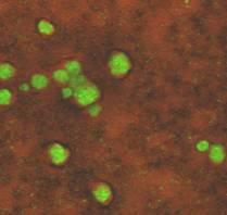
The following movie, constructed with the help of computer software, is from a series of still images recorded through a microscope. It illustrates what can be done with modern microscopes coupled with computer programs that can create movies from still images. This movie demonstrates that some biofilm colonies form in such a manner that channels exist throughout the biofilm. That these channels are open and in fact function as conduits which can deliver oxygen and nutrients deep within the biofilm is shown by this video clip (Video 1).

In this movie, biofilm has been grown in a flow cell with the bulk fluid flowing from right to left. The particles are fluorescent spheres approximately 5 µm in diameter. Their rapid movement indicates that the deep channels in the biofilm are in contact with the bulk fluid layer many micrometers above.
Brightfield and Electron Microscopy
In the 1970s and 80s options for viewing biofilms were chiefly brightfield and electron microscopy (EM-Chap 8) (See exercise entitled: Flow Through Gram Stain). The use of each of these instruments requires the subject to be dehydrated and typically stained (Molecular probes - Chapter 8). The optical resolution of a brightfield microscope (about 0.25 µm) is too small to reveal much detail and although the detail seen under electron microscopy was magnificent, it was misleading. Eighty five to ninety five percent of a biofilm consists of water; the rest is cells, inorganics, and various polymeric materials that are often quite fibrous in nature. The removal of the water, necessary in electron microscopy, results in an image of bacterial cells lying within a flat matrix of fibers. Trying to intuit the nature of a biofilm from electron micrographs is roughly equivalent to deducing the nature of an animal from a long dead road kill.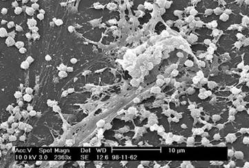
Confocal Microscopy
Perhaps the single most significant advancement in microscopy that permits a more realistic view of microscopic biofilm structure is the confocal microscope (see Confocal Microscopy - Chapter 8). Confocal microscopy involves lasers and computer image processing software that permit observation of a wet subject on virtually any sort of surface.
Without going into great detail, lasers and mirrors are used to produce a series of images of a biofilm, each taken at a slightly different depth inside the biofilm. These image slices allow scientists to view the internal structure of the biofilm. Computer software can in turn be used to create a composite view of the biofilm from the slices, and rotate it to allow one to see the biofilm from the side rather than the top.
Old man van Leewenhoek would be envious.
(See exercise entitled: Building a Biofilm Model from a Confocal Microscope Stack).