Building a Biofilm Model from a Confocal Microscope Stack
Instructor Version (Student Version)
| Subject Area(s) | microbiology |
| Intended Audience |
high school biology, independent study/science fair, introductory undergraduate microbiology, advanced college level microbiology |
| Type | laboratory exercise |
| Revision Date | July 2, 2004 |
CONTENT
This exercise describes the construction of a biofilm model using data obtained from a mixed biofilm of Pseudomonas aeruginosa, P. fluorescens and Klebsiella pneumoniae.
PREREQUISITES
Students should be able to define a biofilm, describe the differences between biofilm (surface-attached) and planktonic (suspended bacterial cells) bacteria, and describe HOW bacteria tend to grow on surfaces.
INSTRUCTIONAL OBJECTIVE
Given readily accessible materials, and the image series associated with this exercise, the instructor or the student will be able to make a biofilm model from foam board, Styrofoam or other similar material that will illustrate the basic structure of a biofilm.
INSTRUCTIONAL PROCEDURES
Introduction
A finished and working reactor system will be shown to the students.
A Confocal Scanning Laser Microscope is to optics what an MRI is to x-rays. It is a microscope that can record a visual slice of heavily hydrated material attached to a surface. Biofilms are such materials and the introduction of the CSLM has revolutionized our view of the structure and complexity of biofilms. A CSLM records an image of a thin slice of a biofilm at a particular focal point, the substrate surface for example, and stores this image in a computer. The instrument then moves to another image plane and records another image. This process is repeated from the substrate surface until the top of the biofilm is reached. The computer can then manipulate this series of images to produce xy (length and width) xz (length and depth) or three-dimensional images. This sort of image processing software is not commonly available in an undergraduate laboratory. Nevertheless, using those same images, one can produce a three-dimensional model which students can use to understand biofilm complexity and structure.
Background
- Accompanying this exercise is a series of confocal images of a mixed biofilm courtesy of Dr. Paul Stoodley, produced at the Center for Biofilm Engineering, Montana State University, Bozeman. See ATTACHMENTS
- Each frame is derived from an image captured by a CSLM at a particular depth into the biofilm. Frame 1, for example, represents the biofilm from the substrate surface to a height of 5 µl above the substrate surface. The second image records the biofilm structure from 5 µl to 10 µl above the surface and so forth.
Building the Model
- The model may be constructed by the instructor, a student or an entire class.
- Photocopy the images and staple or glue them individually onto sheets of foam board, Styrofoam or other easily worked material. These images will serve as your template.
- Using a jig saw, coping saw or modeling saw, cut the dark biofilm sections out of the foamboard. Using the original image as a guide, place the sections of the first image on a plastic, cardboard or wooden base. Glue them into place.
- Repeat this process with each successive layer until the entire biofilm (10 layers) has been built up (Figure 1). Allow the glue to dry thoroughly.
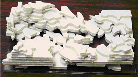
Figure 1. Biofilm Model
MATERIALS AND EQUIPMENT
Quantity |
Description |
| 2 | sets of confocal images (see Attachments below) |
| NA | white glue or other type of glue |
| 1 | wood, plastic or cardboard base (approximately 12 x 20 cm) |
| As Necessary | sheets of 1/4 inch foam board, Styrofoam, or some other material from which the model can be constructed |
| 1 | coping saw, model saw or jig saw |
| As Necessary | safety goggles |
ASSESSMENT / EVALUATION
The student should be able to describe/determine:
- the structural complexity of a typical biofilm;
- that the biofilm is far from a uniform layer;
- if there is more biofilm or bulk fluid volume in the biofilm;
- in which direction the fluid was flowing. Hint: the streamers point down stream;
- what factors might determine the shape of a biofilm.
ATTACHMENTS
- The series of 10 confocal microscope images
- A printable pdf file of the 10 confocal microscope images
REFERENCES
Confocal laser scanning microscopy for analysis of microbial biofilms. Lawrence JR, Neu TR. Methods in Enzymology 1999; 310:11-144
Educational Program Curricula and Teaching Resources
Supported in part by the Waksman Foundation for Microbiology
Developed in collaboration with Dr. John Lennox, Penn State University-Altoona
© 1999-2008 Center for Biofilm Engineering, http://www.cbe.montana.edu